Donnerstag, 17. November 2016

HIV

How is HIV transmitted?
  • 1)     Contact with blood of HIV infected person through cuts in the skin (also through shared injection needles
  • 2)      Breast milk
  • 3)      Semen and vaginal fluids

History of the disease:
A person gets infected with the HIV virus. 1-2 months after infection, the person gets an infection with fever, headache or rushes. Afterwards, the HIV can stay dormant inside host cells for many years. After around ten years, the immune system is so weak that small infection can already be life-threatening. This stage is called AIDS, when the HIV viruses have killed so many CD4 host cells that there are less than 200 CD4 cells per mm^3.
Basic structure:


Life cycle:
At first, a person is infected either via another person that is infected with HIV.  The HIV virus searches for a host cell in the human blood. This is the CD4 cell, a white blood cell. The HIV virus goes into the the host cell by docking on to a receptor and using endocytosis to enter the cell. There, by reverse transcriptase, it converts its RNA into DNA, which can then enter the nucleus. In the nucleus, the DNA of the virus inserts itself into the cells DNA and thus is transcribed and translated. Thus, proteins are produced for new HIV in the cell. The newly created cells, together with HIV RNA exit the cell by budding of and form new HIV cells.

The effects of HIV on the immune system:
The HIV virus kills the white blood cells, called CD4 cells, which help to protect the body from diseases. Through the progressing destruction of these CD4 cells, the immune system becomes weaker and weaker. When infected, the person will develop a small infection after 1-2 months. In the following years, the HIV can be passive and be dormant in the host cell. After 10 years, the immune system is so weak that a small infection can kill the person.

Why can the body not get rid of HIV?
Because the HIV takes a host cell to replicate, because it does not have an own metabolism. It takes the CD4 cell to replicate inside its DNA. This means, that antiviral drugs would have to aim to kill these CD4 cells, to kill HIV, but through this they would destroy the majorly important CD4 cells, which are part of the immunity of the body.

Why does nobody die directly after an infection with HIV?
Because at the beginning, the HIV has to replicate, because there are too little viral cells to be able o cause much damage to the immune system. Thus, as soon as the HIV viruses enter the body, they will hide in the host cell. Throughout the course of the 10 years of infection, the viruses will slowly destroy their host cells, which can then not anymore defend the body from the HIV virus. Thus, after 10yars the body is finally to weak to fight against HIV.

Opportunistic infections which can lead to death in weakened immune systems:
Salmonella, Pneumonia, bronchitis

Cancers:
Invasive cervical cancer, non-hodkin lymphoma, anal-, liver and lung cancer

Other symptoms:
Fever, chills, rush, mouth ulcers, night sweats, swollen lymph nodes, muscle ache, sore throat

Tests:
A laboratory test, whichtests for HIV antibodies and antigens in the blood is conducted. There is a window period in the first 4 weeks, where you cannot detect HIV infection in your blood, but after 6-8 weeks the reliability of the test increases.

Treatments:
In different stages, different antiretroviral therapy(ART) is done. A combination of 2 are taken in each stage (called antiviral regimen). The drugs are also called antiretrovirals (ARV):
For stage 1: Entry inhibitors (before virus enters host)
Stage 2: Fusion inhibitors (stop virus from being docking on to receptor)
Stage 3: non-nucleoside reverse transcriptase inhibitors, nucleoside reverse transcriptase inhibitors (stopping virus from converting into DNA)
Stage 4: Integrase inhibitors (Stopping virus from joining cell DNA)
Stage 5: protease inhibitors (stopping viral proteins from being produced)

Vaccination:
There is no vaccination on the market yet but scientists are working on it.

Prevention:
Getting tested
Know the sex partner’s HIV status
Use condoms
Don’t inject drugs
Take pre-exposure prophylaxis

Social implications of for infected (voluntary or mandatory):
Stigmatization and discrimination of HIV: Many people refer to it in a prejudiced way and have negative attitudes towards people who have HIV. People can be afraid because of fear of contagion. The infected have a low reputation because many people don’t approve of their sex life or habits (drug use). 


Montag, 26. September 2016

Emphysema: Causes, Symptoms and Biological Consequences

The disease causes the air sacs in the lungs, called alveoli, to enlarge. Cell walls are broken down and damaged, resulting in fewer larger damaged air sacs. Thus, the oxygen uptake into the blood and CO2 return is decreased.

Causes:

·         The most important cause is cigarette smoking
·         Air pollution, dust or chemicals
·         Childhood respiratory disorders
·         Low weight

It is caused by an increased level of phagocytes which inflame the wall of alveoli.
The disease can also be congenital (meaning that it is genetically predisposed). People have an alpha1-antitrypsin deficiency.

Symptoms:

·         Shortness of breath
·         Coughing
Minor symptoms:
·         Lung infections
·         Producing mucus
·         Weight loss
·         Fatigue
·         Blue lips and nails
·         Depression
·         Sleep disorder
·         Headache

Biological consequences:

There is an increased level of phagocytes in the alveoli. These normally destroy antigens by engulfing and producing a protein digesting enzyme called elastase, which kills the antigen. An enzyme inhibitor, called alpha 1-antitrypsin normally protects the lung tissue from the elastase by inhibiting the enzyme.
In smokers the number of phagocytes increases, which can therefore inflame the inner wall of the alveoli, because there are not enough inhibitors to inhibit elastase.

Sources:

https://www.google.co.uk/search?q=emphysema&biw=1242&bih=557&source=lnms&tbm=isch&sa=X&ved=0ahUKEwj2qM_g2q3PAhXBvxQKHV_IDi0Q_AUIBigB#imgrc=yQfTRFNQF7dfpM%3A


Lung Cancer: Causes, Symptoms, Biological Consequences

Cancer generally can be defined as out-of-control cell growth.
There are 2 types of tumour, the malignant and the benign tumour. The malignant tumour can spread to other organs of the body, causing secondary tumours to arise. This is more dangerous than a benign tumour which stays at its original location. The process by which a primary tumour is spread in the body to form secondary tumours is called metastasis.

Causes:

Inhalation of carcinogenic substances (x-rays, tobacco, air pollution, chemicals, dust). These are responsible for damaging the DNA. Free radicals form, which want to steal electrons from other body cells and in the process they damage the DNA. Humans can also be born with a predisposition to develop cancer due to a genetic mutation which makes cancer more likely.

Symptoms:

The cancer undermines the lung’s ability to take up oxygen. It tends to block air way paths and tubules. There can also be bleedings. Another sign might be chronic coughing.
  • ·         Coughing intensely
  • ·         Pain in shoulder or chest
  • ·         Changes in coughed up mucus
  • ·         Coughing blood
  • ·         Chronic pneumonia
  • ·         Chronic bronchitis
  • ·         Difficulty breathing
  • ·         Hoarse voice


Biological consequences:

Cancer can occur in 2 situations: either it can spread through the blood by a process called invasion, destroying healthy tissue, or a cell divides and grows and feeds on blood by a process called angiogenesis.

There is small cell lung cancer (SCLC) and non-small cell lung cancer (NSCLC)

Examples of NSCLC:

  • Squamous cell carcinoma:
  • ·         Most common
  • ·         Most common in men
  • ·         Forms in lining of the bronchial tubes

  • Ardenocarcinoma:
  • ·         Most common in non-smokers
  • ·         Most common in women
  • ·         Forms in mucus producing glands in the lungs


Sources:
https://www.google.co.uk/search?q=squamous+cell+carcinoma&espv=2&biw=1242&bih=602&source=lnms&tbm=isch&sa=X&ved=0ahUKEwit_YCJ2K3PAhVFKsAKHVc9BU0Q_AUIBigB#imgrc=MLiJCKzMn8aLuM%3A
https://www.google.co.uk/search?q=Adenocarcinoma&espv=2&biw=1242&bih=602&source=lnms&tbm=isch&sa=X&ved=0ahUKEwizqYyP2K3PAhVqI8AKHR76B6cQ_AUIBigB#imgrc=J2z4VMXs7e_2OM%3A


Sonntag, 25. September 2016

Olaf the oxygen malecule and his journey through the human body

Hello I am Olaf, the oxygen molecule and I want to take you around the body and show you my daily life, supporting the human respiratory system...

You can imagine my cycle around the body like a day at work, starting in the morning when I go to my work place. My state from floating around in the air changes because person x inhales my through his mouth. I am sucked into the trachea then, which is like like walking along a street.

This happens because of the change of pressure in the internal body system and the external pressure. The diaphragm moves downwards pushing the abdomen out and the rib cage moves upwards and outwards at the same time, so that the lungs have more space. Thus, there is an increased volume which decreases the pressure inside the lungs, causing the air to be inspired.

So I travel down the pathway from trachea to the lungs, starting with big tubes called bronchi, which separate into smaller tubes called bronchioles which end in little air sacs, called alveoli.

Arrived at the alveoli, now I have to cross the membranes to get to the blood stream, flowing through a network of little blood vessels called capillaries. This is like a crossing over a pedestrian crossing. I have to float through two cell walls, one of the alveoli and one of the capillary, having each two membranes, thus until arriving in the bloodstream crossing a total of four cell membranes, which is a relatively short way for me, making my day less complicated.

Now, I jump onto a red blood cell which will carry me through the stream, like a personal motor boat. Meanwhile I oxygenate the deoxygenated stream. Special about it is, that it is an artery stream, which I travel along in direction of the heart now, which can be imagined like travelling in the opposite direction of the traffic, because normally arteries lead away from the heart. To jump on the boat and to be bound to a hemoglobin molecule, I have to overcome the wall of the red blood cell, which is like opening the door of a car.

The red blood cell takes me to the heart, which is like a junction, where the direction of traffic changes. Now I am pumped together with the red blood cell into a new artery, which brings me to whatever location where engergy is needed. Arrived at any place in the body, I can mix with my colleagues, a few Mr Water and a particular Mr Glucose. Together, we can produce ATP.

This is basically the important bit of my work and afterwards I am really exhaused and I head of into direction of the outside world. Thus, another red blood cell picks me up via a vein. I have to cross its cell wall again then, binding to a carbohemoglobin that is connected to the cell. On the way back, we cross the juction at the heart again leading to another vein that belongs to the pulmonary system, which my day had started with, the circulation to the cells for energy having been the systematic circulation.

The vein takes me back to the lungs where I have to at first get off the blood cell by exiting through its membrane and then cross the four membranes of capillary and alveoli again, ending in the alveoli as a carbon dioxide compound.

Now, the only step that is left, before my final destination, returning to my home, the air, is to be pumped up by exhalation of person x again. Ventilation is activated by the diaphragm, a muscle which relaxes and pushes upward into a domed shape and the rib cage being pulled in and down. I am moves outside through the bronchioles, bronchi and trachea again, finally floating through the mouth and being released into the atmosphere.

This was my daily routine and the formation of O2 into CO2 in the human respiratory system.

Sources:
http://de.slideshare.net/sarahfree/oxygen-through-the-respiratory-system
https://prezi.com/9uqpc_zz4x1l/oxy-the-oxygen-molecules-journey-through-the-respiratory-system/
http://image.slidesharecdn.com/lecture7resp-141208082139-conversion-gate01/95/respiratory-system-7-638.jpg?cb=1418029376


Samstag, 17. September 2016

COELIAC DISEASE - WHAT IS IT ABOUT?

What celiac disease is:

The lining of the small intestine after the immune system has attacked the gluten
It is an autoimmune disorder. This means that the body attacks its own tissue. This results in inflammation of organs which can be really dangerous. Celiac disease affects the gastrointestinal tract, because the body recognizes gluten, found in food such as barley, rye and wheat as a threat to the body and attacks it. While attacking the gluten, the villi in the small intestine get also damaged through the inflammation and are shortened, which means that they decrease in surface area. This means that they can’t absorb nutrients as efficiently anymore and this results in nutrient deficiencies and so on.

Description of the basic cause and symptoms of celiac disease:

Celiac disease is caused by the body’s own immune system, which reacts and attacks the gluten found in the gut. The disease can be caused partly environmentally and partly by genetics and occurs more often in people who already suffer from another disease. It can develop spontaneously and can be unnoticed by the person who has got it. The villi in the small intestine are damaged and thus nutrients can be absorbed properly anymore. This damage in the intestine leads to symptoms such as
·         weight loss due to malnutrition caused by the poor absorption.
·         diarrhoea and abdominal pain and swelling due to the inflammation.
·         food intolerance
·         inflammation increases the risk of gastrointestinal cancers (e. g. cancer of oesophagus or small intestine)
·         iron deficiency due to lack of red blood cells
·         vitamin deficiencies
·         low bone mineral density
·         visible affects: enamel of teeth, chronic fatigue, joint pain, poor growth, delayed puberty, miscarriage and infertility
·         Neurological symptoms: Migraine, depression, ADHD, epilepsy

Explanation of the effect of celiac disease on intestinal villus and how this leads to its symptoms:

The villus is inflamed and thus shortened and flattened so that nutrients cannot pass through it anymore. This causes the cells to be more sensitive so that cancer can be developed. It also means that nutrients such as vitamins cannot be taken up into the blood, so that vitamin 12 and folate deficiency anaemia can be developed. Iron deficiency also causes a lack of red blood cells, leading to other health problems, connected to oxygen transport.

Description of the effect of genetics on a person’s predisposition to celiac disease:

There is a genetic predisposition for the development of the disease and first degree relatives have a 4-15 % chance to inherite the disease, but the inheritance pattern is unknown. The disease seems tobe connected to the disposition of the genetic of a human containing specific variants of the HLA-DQA1 and the HLA-DQB1 genes. These are human leukocyte antigen complexes (HLA). They bond by proteins called antigen-binding DQaß heterodimer, which is a complex that is on the surface of immune cells and attaches to antibodies, which it recognizes as invading.

How celiac disease is currently tested for:

Australian scientists found a method to test the blood for celiac disease in 2014. Generally, people who are genetically predisposed to have the disease are advised to be screened. The diagnosing nowadays involves the taking of the blood sample to search for antibodies that are specific to that disease. When they are found, a biopsy is conducted whereby an endoscope(a tube with light) is passed down the mouth to the small intestine and  a tiny biopsy tool will take samples of the small intestine.

Sources:

https://www.google.co.uk/search?q=picture+of+coeliac+disease&espv=2&biw=1242&bih=606&source=lnms&tbm=isch&sa=X&ved=0ahUKEwiBtsT-iZfPAhUjI8AKHcgnBggQ_AUIBigB#imgrc=ypRXZzd0mYwyCM%3A


TESTING A TRANSPIROMETER

A transpirometer is there for the purpose of measuring plant transpiration. Transpiration can be defined as 'the passage of water through a plant from the roots through the vascular system to the atmosphere'.
The instrument constists of a tube with 3 openings(in one of which the leavy shoots sits). A horizontal thin gradualted cappilary tube is connected to it's horizontal end and leads into a beaker. The beaker controls the constant level of water in the tubes and a reservoir is connected to the system to be able to reset the experiment. The tubes are all filled with water, allowing just one little bubble of air into the capillary tube and sealing the opening to the leavy shoot with a cork and vaseline so that no additional air can come into the system. When the plant transpires, water is supposed to be sucked up by the roots, so that the air bubble will start moving along the graduated caillary tube. The scaling bars on it allow to measure the rate of transpiration.

In my biology class we conducted a set up the transpirometer, the photos of the set up are shown below:


http://c1.staticflickr.com/1/458/20406535075_f2432a61b2_n.jpg

http://www.merriam-webster.com/dictionary/transpirometer

Donnerstag, 14. April 2016

About the development in science in the field of human cloning

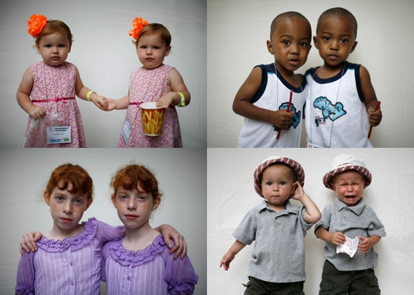
Identical twins http://www.slate.com/content/dam/slate/articles/health_and_
science/science/2014/09/140912_SCI_IdenticalTwins.jpg.CROP.
promo-mediumlarge.jpg

Human cloning has recently acaused a sensation in science, because of a Chinese scientist, who is working in an animal cloning factory, who announced, that his department is ready to clone humans now after having taken the step with other animals successfully. I read this in the following article:
https://www.rt.com/news/324110-china-human-cloning-factory/
In the article is said that the technologies are ready to clone humans, which I think sounds very frightening, because as in many other articles mentioned, there could be loads of ethical issues, which would describe, how dangerous the cloning itself could be for the cloned individual and in general in is frightening to think about how dangerous it could be if humans like Hitler would be brought back to life. In the article is also talk of how infertile couples could have the chance for a child, identical to one parent, but seriously, how would that freak people out to have to see one's exact version walking around, I think it would drive people crazy in a very negative way.
I also watched a video, which can be found in the following like, which was very informative.
https://www.youtube.com/watch?v=Tz8HxNfIG8Q
Here is talked about the beginning in the scientific research about the general cloning of animals and I wonder here slightly, why not the person, who initially cloned the first animal in gerneral, the guy mentioned in the video called John Gurdon, is honored more for his findings, by cloning a frog, while everybody celebrates Wilmut and Campbell, who cloned Dolly the sheep, although this happened far later in 1996.
What I found the most interesting though in the video was the mention of the fact, that scientist called Robl and Cibelli claimed in 1996 to have successfully transplanted a human cell of Cibelli's leg and cheek cells into an egg cell of a cow. To find out more information about this I researched them and found this article 
http://www.nytimes.com/1998/11/12/us/researchers-claim-embryonic-cell-mix-of-human-and-cow.html?pagewanted=all
If these scientists tell the truth and have really conducted a research with that outcome, this would be very scary indeed because it would mean that even with rather simple equipment they were able to conduct their research. Thus, I think scientists should be careful in the future to ban human cloning very strictly and under control, because in my opinion it is not ethically right to do such things and there could just be loads of disadvantages and risks such as a higher risk for diseases throgu the minimising of variation in human genes.